The cochlea is shaped like a snail--a long tube which is wound around in a spiral. On the big end (where the snail head would be), it has two soft spots, the oval window and the round window, which both open onto the air cavity of the middle ear. The stapes touches and pushes on the oval window, but nothing pushes on the round window. The cochlea is embedded in the bone of the skull.
The fact that the cochlea is wound up is similar to a tuba or certain other wind instruments being wound up--it is simply a way for a long, thin tube to fit inside a smaller space. The full length, uncoiled, of the cochlea would be about 3.5 cm, whereas its actual diameter is about 1 cm. It is easier to picture how the cochlea works if we think about the unwound version, so from here on we will think about it as a long, narrow tube, round in cross-section and slightly tapered towards the point.
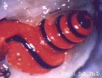
The cochlea's tube is divided by two membranes into three fluid-filled regions running the length of the cochlea. Here is a creepy photograph of a cochlea in which two of the regions are filled with a red gel and the third is filled with a blue gel:
(The nice looking figures were ruthlessly copied from A webpage about hearing which is also worth looking at. The last figure is from another nice site.)
Here is a cross-section (actual photograph) of what you would see if you cut the tube of the cochlea open, that is, a cross-section of the tube:
The top region is called the Scalar Vestibuli. A membrane (Reissner's membrane) separates it from the Scala Media, which is separated from the Scala Tympani by a membrane (the basilar membrane) which carries a gelly-like body on its top, called the organ of Corti.
So here is a bad cartoon of the cochlea, as it would look if it were unwound. The oval window touches the Scala Vestibuli and the round window touches the Scala Tympani. The two are connected at the back by a tiny hole called the heliocotrema.
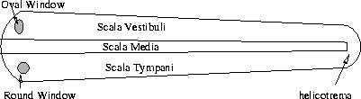
When the stapes pushes in or pulls out the oval window, it squishes or stretches the fluid in the Scala Vestibuli. That fluid has to go somewhere. The whole cochlea is embedded in bone, except the round window. Therefore, the fluid has to cause the round window to bulge out. How can it?
There are two ways, the ``fast way'' and the ``easy way.'' The ``fast way'' is for the extra fluid in the Scala Vestibuli to ``dent in'' the membranes between the 3 chambers, squishing the Scala Tympani, and squishing out the round window:
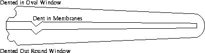
The ``easy way'' is for the fluid to run the length of the cochlea, through the helicotrema, and back.
The trick is that the membranes across the cochlea are stiff near the beginning and get softer and softer as you go the length of the cochlea. Therefore, it is faster to make the dent near the beginning, but easier to make it near the end. The faster you pull back and forth the oval window, the nearer the beginning of the cochlea the ``dent'' will appear, because there is not time for the fluid to go further. The slower you pull back and forth the oval window, the nearer the end the ``dent'' will appear, because there is time for the fluid to go to where the membrane is more flexible. Therefore the location, along the cochlea, where the membrane ``dents in'' will vary according to how fast the oval window is being pulled in and out. We will see that this is one of the two ways that we can tell the pitch of a sound.
We still have to see how that motion is converted into a nerve impulse. To understand that, we need to look closely at the organ of corti, on the membrane between the Scala Media and the Scala Tympani. We just saw that, when there is a sound, these membranes will get squished (or stretched, if the oval window gets pulled out by the stapes).
Here is a zoom-in of the cross-section of the Organ of Corti:
You see that there is a membrane hanging into the Scala Media, called the Tectoral Membrane. It separates a little bit of the Scala Media from the rest. When the Scala Media is pushed on from above, the tectoral membrane moves back and forth with respect to the membrane below it, essentially because it is "tied to the wall" while the body below it is "tied to the floor". The mass of tissue underneath that channel contains rows of cells (rows stretching along the length of the cochlea, so when you look at the cross-section you see just one cell), called hair cells. Each cell has about 100 stereocilia, which are like tiny hairs, projecting off its top into the fluid of the channel. They either touch, or are actually anchored in, the tectoral membrane. When the tectoral and basilar membrane move with respect to each other, it pulls these cilia (hairs) back and forth. The hair cells are nerve sensors. That means that the back end of the cell is attached to nerve cells, and when the hairs move, it triggers an electrical impulse to be sent to the nerve cells it attaches to. So the order of cause and effect is,
- Sound--pressure--on the eardrum pushes the ossicles, which push the oval window;
- Fluid in the Scala Vestibuli flows partway along the cochlea, where it pushes in the Reissner's Membrane and makes the fluid in the Scala Media flow;
- Fluid in the Scala Media moves, stretching the basilar and tectoral membranes in different directions;
- Shearing motion between the membranes pushes back and forth the stereocilia (hairs) on the hair cells;
- Hair cells respond by sending an electical signal to the nerve cells, which will communicate it down nerves to the brain.
It is a subject of future lectures, how the properties of the sound (pitch, loudness, timbre) are related to the ways they stimulate the hair cells and send a signal to the brain.